0% interest EMI options · Clear treatment costs explained before you begin · Book consultation

Hysteroscopy treatment at PRAVI IVF
A closer look inside the uterus when there is a clear reason to investigate.

Hysteroscopy in Kanpur

Hysteroscopy allows a doctor to examine the uterine cavity using a thin camera passed through the cervix. It may help investigate abnormal bleeding, suspected polyps, scar tissue or certain uterine abnormalities that could affect fertility. Some findings can be treated during the procedure, but this is not always necessary or possible. At PRAVI IVF in Kanpur, consultation can help clarify whether hysteroscopy is appropriate and what pathway is available.

  • Hysteroscopy examines the inside of the uterus; it does not directly assess the ovaries or the outside of the uterus.
  • Diagnostic examination and operative treatment have different preparation, pain relief and recovery requirements.
  • Hysteroscopy is not a routine requirement before every IVF cycle or after every unsuccessful embryo transfer.

Who this is for

  • People whose ultrasound suggests an endometrial polyp, a cavity-distorting fibroid or another finding needing closer assessment.
  • People with persistent abnormal uterine bleeding when examination of the cavity or a tissue sample is indicated.
  • People with suspected intrauterine adhesions, particularly after uterine procedures or with a change in menstrual flow.
  • People being evaluated for recurrent pregnancy loss when a uterine cavity abnormality is suspected.
  • People with unsuccessful embryo transfers who have a specific reason for further uterine assessment, rather than a routine need for hysteroscopy.

Why patients choose PRAVI

High

IVF success rate

6,000+

Babies delivered

4.9/5

600+ patient reviews

18+

Years of fertility care

Mon – Sat · 9:00 AM – 7:00 PM

Step by step

How the Hysteroscopy process works

01

Establish the reason for examination

The clinician reviews bleeding patterns, fertility history, previous uterine procedures and scan findings. This helps determine whether hysteroscopy could answer a specific question or whether imaging or another investigation should come first.

02

Agree on the scope and consent

Discuss whether the plan is examination alone, biopsy or possible treatment. Pain relief, anaesthesia, alternatives and consent limits should be explained, including what happens if an unexpected finding requires a separate procedure.

03

Examine the uterine cavity

A thin hysteroscope is passed through the vagina and cervix into the uterus. Sterile fluid gently opens the cavity for viewing; a tissue sample or targeted treatment may follow if planned and appropriate.

04

Review recovery and findings

Afterwards, the team assesses symptoms and recovery before giving discharge advice. Findings, any tissue results and the need for further treatment are reviewed before deciding when fertility treatment or attempts at pregnancy can resume.

What hysteroscopy can reveal inside the uterus

Hysteroscopy is a direct examination of the endometrial cavity, the space inside the uterus where a pregnancy implants. A narrow telescope with a light and camera passes through the vagina and cervix, so there is no abdominal incision. Sterile fluid separates the uterine walls, allowing the clinician to inspect the lining, cavity shape and openings of the fallopian tubes. Seeing these openings does not establish whether the tubes are open along their full length.

The examination may identify polyps, fibroids projecting into the cavity, adhesions or a suspected septum. A biopsy can be taken when the lining needs laboratory assessment. Diagnostic hysteroscopy means looking and sometimes sampling; operative hysteroscopy involves treatment with instruments. Neither automatically includes the other. Hysteroscopy cannot fully assess the outer shape of the uterus, diagnose all causes of infertility or explain every unsuccessful embryo transfer, so findings need to be interpreted alongside other investigations.

When hysteroscopy may help, and when it may not

A clear clinical question is the genuine starting point. Hysteroscopy may be useful when ultrasound suggests a cavity abnormality, bleeding remains unexplained or adhesions are suspected after a uterine procedure. It can also form part of an individual assessment for recurrent pregnancy loss. Whether treatment of a finding is worthwhile depends on its size, position, symptoms and relevance to the person's reproductive history, not simply on its presence.

An unsuccessful embryo transfer does not by itself prove that something inside the uterus needs correcting. Routine hysteroscopy is not necessary for everyone with infertility, particularly when appropriate imaging is reassuring and there are no concerning symptoms. It will not address sperm problems, reduced egg supply or every cause of implantation difficulty. The decision should compare the likely value of information or treatment against discomfort, procedural risks and a possible delay in fertility care.

  • Ask which specific finding or symptom the procedure is intended to investigate.
  • Ask whether the result is likely to change your next treatment decision.

Assessment before deciding on hysteroscopy

The assessment usually begins with menstrual and bleeding history, previous pregnancies, fertility treatment and any uterine surgery or infection. Bring earlier ultrasound reports, procedure notes and available tissue results. A transvaginal ultrasound is often the initial examination. Depending on the question, saline infusion ultrasound or three-dimensional ultrasound may provide useful detail without immediately proceeding to hysteroscopy. Suspected uterine shape abnormalities sometimes need additional imaging because the external contour affects diagnosis and treatment choices.

Before booking, the clinician should consider pregnancy possibility, infection symptoms, medicines and conditions relevant to bleeding or anaesthesia. Hysteroscopy is generally avoided during pregnancy and postponed with active pelvic infection. Timing is often chosen when the lining is relatively thin, but irregular cycles and urgent bleeding concerns may require a different approach. Not everyone needs the same blood tests or pre-anaesthetic assessment. Ask what testing is necessary for your planned procedure and why.

How examination and targeted treatment are performed

Diagnostic hysteroscopy may be performed in an outpatient setting with an agreed pain relief plan. Other procedures require sedation or anaesthesia in an operating theatre, particularly when more extensive treatment is expected. The cervix may need gentle widening before instruments can pass. Once the cavity is visible, the clinician records relevant findings and may take a biopsy. If discomfort is unacceptable or access is difficult, stopping and arranging another approach can be safer than continuing.

Selected polyps or cavity-projecting fibroids can sometimes be removed hysteroscopically. Adhesions may be divided, although dense scarring can be difficult to treat and may recur. Septum surgery requires a confirmed diagnosis and discussion of the uncertain benefit in the individual's circumstances; it is not an automatic response to any unusual cavity shape. Larger or complex findings may need staged treatment or referral. Unexpected findings should be managed within the consent agreed beforehand, rather than treated without discussion.

Risks, discomfort and limits of hysteroscopy

Cramping, pain, light bleeding and feeling faint can occur during or after hysteroscopy. Some people find an outpatient examination quite uncomfortable, so pain relief and the option to stop should be discussed in advance. Infection, heavier bleeding and injury to the cervix are possible. Perforation means an instrument passes through the uterine wall; depending on the circumstances, observation or further surgery may be needed. Sedation and anaesthesia have their own risks, which require a separate assessment.

Operative treatment brings additional considerations, including absorption of the fluid used to view the cavity, injury involving nearby organs and new intrauterine adhesions. Risks vary with the procedure's complexity and individual health. Removal of a finding does not guarantee improved implantation or live birth, and some abnormalities need repeat assessment or treatment. Hysteroscopy does not involve operating on the ovaries and is distinct from laparoscopy or ovarian drilling. Any additional procedure needs its own indication and consent.

Preparing for the planned type of hysteroscopy

Preparation depends on whether the visit is for an awake diagnostic examination or operative treatment with sedation or anaesthesia. Confirm the setting, expected scope and pain relief options before attending. Do not assume fasting is required for every hysteroscopy; follow the instructions provided for your procedure. Tell the team about allergies, previous anaesthetic problems and all medicines, including blood thinners and supplements. Do not stop prescribed treatment without advice from the responsible clinician.

Discuss how pregnancy will be excluded and whether you should avoid unprotected intercourse beforehand. Report fever, unusual discharge, significant pelvic pain or a new possibility of pregnancy before the appointment. If sedation or anaesthesia is planned, arrange an accompanying adult and follow the team's transport and supervision instructions. Bring sanitary pads and allow flexibility for recovery rather than assuming you can return immediately to work. You can also discuss anxiety, previous painful examinations and preferences about support during care.

  • Confirm fasting, medication and transport instructions for your particular procedure.
  • Ask what treatment is authorised if the examination reveals an unexpected abnormality.

Recovery, warning symptoms and reviewing results

Mild cramps and light spotting may follow hysteroscopy, but recovery differs between a simple examination and more extensive surgery. Discharge depends on the procedure and your condition; it should not be assumed to occur the same day in every case. Follow the written advice on pain relief, activity, intercourse and vaginal products. Seek urgent medical assessment for severe or worsening abdominal pain, heavy bleeding, fever, foul-smelling discharge, fainting or feeling markedly unwell. Use the contact instructions provided by the treating team.

Visual findings may be discussed after the examination, while biopsy results require later review. Ask for a clear explanation of what was seen, what was treated and whether anything remains uncertain. Following adhesion treatment or other cavity surgery, a repeat assessment may be considered before pregnancy attempts or embryo transfer. There is no single waiting interval suitable for everyone. The next step depends on healing, tissue results, the original indication and the wider fertility plan.

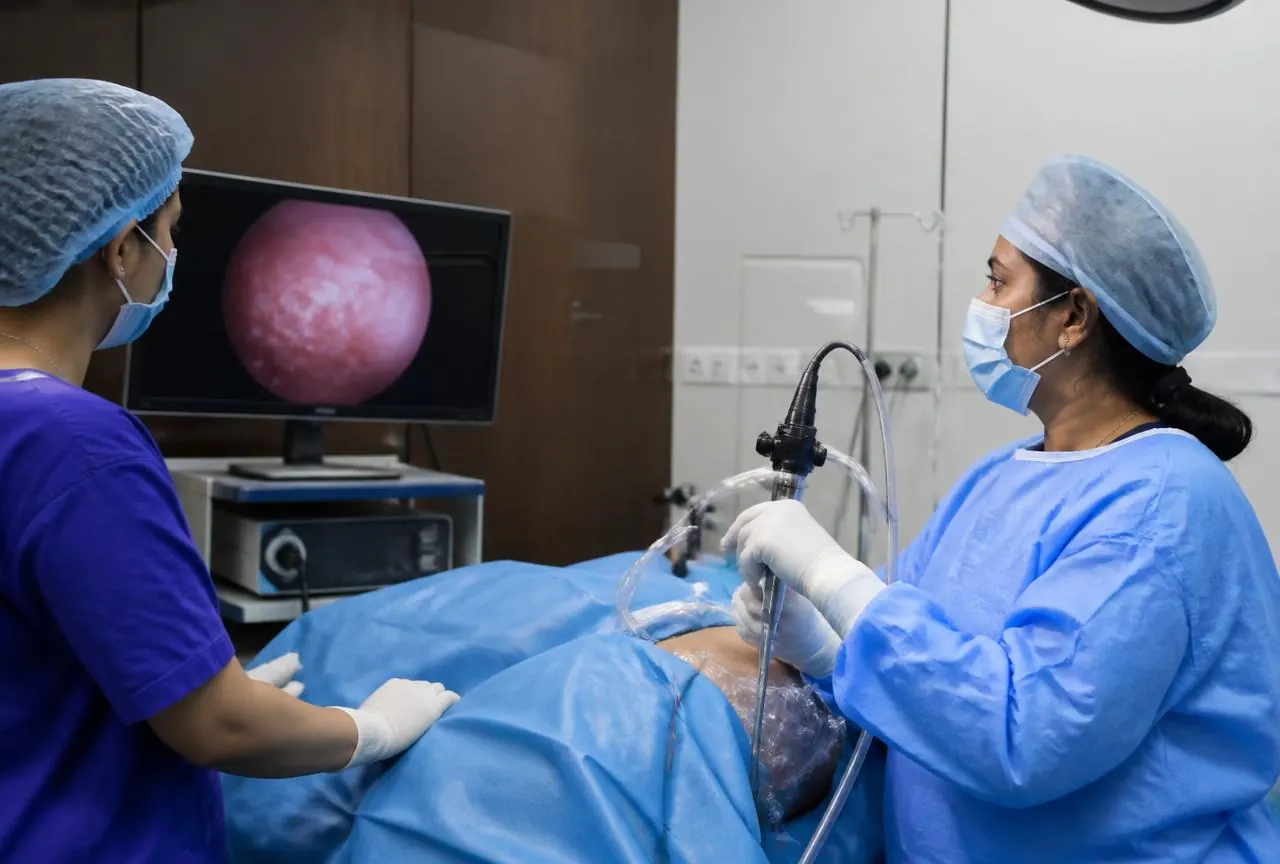
Discussing hysteroscopy at PRAVI IVF in Kanpur

A consultation at PRAVI IVF in Kanpur can focus on whether examination of the uterine cavity is likely to change your care. Bring scans, previous hysteroscopy or surgery reports, pregnancy records and details of embryo transfers where relevant. Ask the clinician to distinguish a suspected finding from a confirmed diagnosis, and a potentially useful intervention from a necessary one. If the recommendation follows unsuccessful fertility treatment, request an explanation of why hysteroscopy is being considered in your particular situation.

Confirm the actual PRAVI pathway, including availability of diagnostic or operative hysteroscopy, the proposed setting, who will perform the procedure and how anaesthesia is arranged if needed. Ask how complex findings, tissue testing and follow-up are handled, and which circumstances could require referral. The consultation should also establish consent limits, alternatives and whom to contact with problems afterwards. The aim is an informed decision about a specific uterine concern, not a promise that surgery will produce a pregnancy.

Frequently asked questions

This information does not replace an individual medical assessment. Treatment suitability, available services and any legal requirements must be confirmed with the clinic before you begin.

Other services