0% interest EMI options · Clear treatment costs explained before you begin · Book consultation

Follicular monitoring treatment at PRAVI IVF
Understanding follicle growth and the timing of ovulation

Follicular monitoring in Kanpur

Follicular monitoring uses a series of ultrasound scans to follow developing follicles in the ovaries and assess the uterine lining. It can help clinicians plan intercourse, insemination or parts of an IVF cycle, but it cannot confirm egg quality or promise pregnancy. At a PRAVI IVF consultation in Kanpur, you can discuss whether monitoring would answer a useful question in your fertility care and how your appointments would be planned.

  • Ultrasound tracks fluid-filled follicles; it does not directly show the eggs inside them.
  • Monitoring can guide fertility-treatment decisions but cannot establish egg quality or guarantee conception.
  • Scan timing and frequency depend on the cycle, medication response and clinical purpose.

Who this is for

  • People with irregular cycles when a clinician needs more information about follicle development and possible ovulation.
  • People taking ovulation-induction medicines who need their ovarian response checked.
  • Couples planning timed intercourse or intrauterine insemination when ultrasound findings may help guide timing.
  • People undergoing IVF who need follicle tracking as part of supervised ovarian stimulation.
  • People being assessed for suspected ovulation problems, alongside other relevant fertility investigations.

Why patients choose PRAVI

High

IVF success rate

6,000+

Babies delivered

4.9/5

600+ patient reviews

18+

Years of fertility care

Mon – Sat · 9:00 AM – 7:00 PM

Step by step

How the Follicular monitoring process works

01

Review the purpose of monitoring

Your clinician reviews cycle patterns, previous investigations and any fertility medicines. Together, you clarify whether scans are intended to assess ovulation, guide intercourse or insemination, or monitor ovarian stimulation during an IVF cycle.

02

Arrange the first ultrasound

An initial scan may assess the ovaries, existing follicles or cysts, and uterine lining. Its timing depends on your cycle and treatment, rather than a fixed calendar date that applies to everyone.

03

Track changes across the cycle

Repeat scans measure developing follicles and changes in the lining. Your clinician decides when another assessment is useful and whether blood tests are needed; monitoring schedules may change according to the findings.

04

Discuss the next clinical decision

Scan findings are interpreted alongside your symptoms and treatment plan. The clinician may advise timing, further observation, medication review or cancellation if the ovarian response makes continuing the planned cycle unsafe or unsuitable.

05

Review the cycle afterwards

Follow-up considers whether ovulation appeared to occur, how the ovaries responded and whether further assessment is needed. If pregnancy is possible, ask when to test and how any prescribed treatment should be continued.

What follicular monitoring can show

A follicle is a small fluid-filled structure in the ovary that may contain an egg. During a menstrual cycle, a group of follicles begins developing, and usually one becomes dominant before ovulation. Follicular monitoring, sometimes called folliculometry, follows this process through ultrasound examinations at selected intervals. The clinician records follicle measurements, their number and appearance, and the thickness and pattern of the endometrium, which is the lining of the uterus. Comparing scans helps show change rather than relying on a single snapshot.

Ultrasound does not show the egg itself, prove that it is mature or assess its genetic health. A follicle that has reduced in size or collapsed, sometimes with fluid behind the uterus, may support an interpretation that ovulation has occurred. These findings still need clinical context. Monitoring helps estimate the fertile window and guide decisions, but it cannot establish whether sperm will fertilise an egg or whether an embryo will implant.

When monitoring may help—and when it may not

Monitoring may be useful when periods are irregular, ovulation is uncertain or medicines are being used to encourage follicle development. People with polycystic ovary syndrome may need scans to distinguish small resting follicles from follicles that are actively growing. During intrauterine insemination, scan findings may help coordinate insemination with expected ovulation. In IVF, monitoring has a different purpose: it helps the team assess the response of several follicles during stimulation and plan later treatment steps.

Regular scans are not necessary for everyone trying to conceive. Someone with predictable cycles and no identified fertility concern may gain little from repeated ultrasound appointments. Monitoring also cannot overcome blocked fallopian tubes, significant sperm problems or other causes of infertility on its own. If pregnancy has not occurred, continuing scan after scan without reviewing the wider picture may delay more useful assessment. Ask what specific decision the result will influence and when the overall plan should be reconsidered.

Assessing your cycle before the first scan

The starting point is a review of your menstrual history, including the first day of your last period, usual cycle length and recent changes in bleeding. Tell the clinician about previous pregnancies, pelvic surgery, known ovarian cysts, endometriosis and any hormonal medicines. A baseline ultrasound may be appropriate, particularly before a medicated cycle, to look at the ovaries and uterine lining. If a period is late or pregnancy is possible, ask whether pregnancy testing is needed before treatment or monitoring proceeds.

Other investigations depend on the question being asked. Thyroid or prolactin testing, selected reproductive hormone tests, semen analysis or assessment of the fallopian tubes may be relevant, but not every person needs every test. Blood-test timing differs between tests and between treatment plans. If anti-Müllerian hormone testing is discussed, remember that AMH mainly helps estimate egg quantity and likely ovarian response; it does not measure egg quality or establish whether natural pregnancy will occur. Previous reports can help avoid unnecessary repetition.

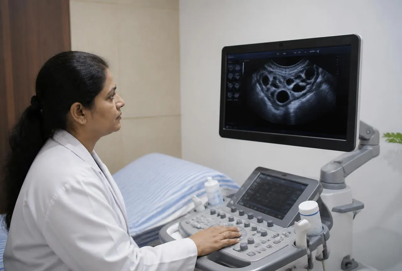
What happens during a follicular scan

Follicular monitoring commonly uses transvaginal ultrasound because it provides a close view of the ovaries and uterine lining. After the procedure is explained and you consent, a covered, lubricated ultrasound probe is gently placed inside the vagina. You may feel pressure or discomfort, especially if you have pelvic tenderness. Tell the person performing the examination if it hurts; you can ask to pause or stop. You can also ask about privacy arrangements and whether a chaperone or support person may be present.

Transabdominal ultrasound, performed over the lower abdomen, may be considered if an internal scan is declined or unsuitable, although it may provide less detail in some circumstances. Bladder instructions differ: an internal scan usually requires an empty bladder, while an abdominal scan may require a full one. Confirm instructions before attending. Ultrasound uses sound waves rather than ionising radiation. The scan measures visible structures; it does not involve collecting an egg, opening the cervix or treating an ovarian cyst.

How scan findings guide timing decisions

There is no single scan day or follicle measurement that reliably predicts ovulation for everyone. The first appointment and the interval between scans depend on your usual cycle, previous findings and whether medicines are being used. Appointments may become closer together as a follicle develops. A clinician may combine ultrasound with urinary ovulation testing or blood hormone measurements when those results would change management. An app's predicted fertile window cannot replace clinical review in a supervised treatment cycle.

For timed intercourse or insemination, the findings can help estimate when an egg may be released. If a trigger injection is part of the plan, its timing and suitability must be decided by the treating clinician; do not use or repeat it independently. In IVF, scans help guide stimulation and egg-retrieval planning rather than intercourse timing. Follicle counts do not equal the number of usable eggs or embryos. Even if fertilisation occurs with IVF or ICSI, it does not guarantee implantation or live birth.

Limits of monitoring and safety during treatment

The ultrasound examination itself is generally low risk, although temporary discomfort can occur. Its main limitations concern interpretation: measurements can vary, ovulation can happen between appointments, and apparently reassuring follicle growth does not exclude other fertility problems. Endometrial thickness is one piece of information, not a pass-or-fail test for pregnancy. A cycle may show no dominant follicle, delayed growth or findings that remain uncertain. These results should prompt explanation and review rather than an automatic increase in treatment.

When fertility medicines are involved, monitoring also helps assess safety. Several developing follicles can increase the chance of a multiple pregnancy if intercourse or insemination proceeds. Excessive ovarian response can also raise concern about ovarian hyperstimulation syndrome, particularly with injectable stimulation. A clinician may advise withholding a trigger, avoiding unprotected intercourse or cancelling treatment. Scans reduce uncertainty but cannot eliminate these risks. Request urgent medical assessment for severe or worsening abdominal pain, marked swelling, persistent vomiting, breathlessness, fainting or reduced urine output.

  • Ask what findings would make the planned cycle unsafe to continue.
  • Clarify who to contact if symptoms develop between appointments.
  • Do not change fertility medicines based only on the written scan report.

Preparing for appointments and reviewing the cycle

Bring earlier ultrasound reports, relevant blood results and a list of medicines or supplements, including any ovulation-induction medicines already taken. Record menstrual dates and follow the appointment-specific bladder instructions. Fasting is not usually needed for follicular ultrasound alone, but check if another test is scheduled. Tell the team about pain with vaginal examinations, a previous difficult examination or concerns about consent before the scan begins. Planning for repeat visits can help, since the next appointment may depend on that day's findings.

Most people can return to usual activities after an uncomplicated scan; sedation and a recovery period are not normally required. The follow-up discussion should explain what changed, what remains uncertain and whether another scan or test is needed. If ovulation was expected but the period does not arrive, follow the clinician's pregnancy-testing advice. Testing too soon after certain trigger injections can be misleading. If the cycle does not lead to pregnancy, review the whole fertility plan rather than assuming that follicle growth alone means everything else is normal.

Discussing follicular monitoring at PRAVI IVF

A consultation at PRAVI IVF in Kanpur can focus on whether follicular monitoring is appropriate for your circumstances and what decisions it is intended to support. PRAVI IVF has centres in Kanpur and New Delhi; confirm the arrangements for your chosen location rather than assuming both follow the same appointment pathway. Exact service availability, scan arrangements, clinician review and consent procedures should be confirmed at consultation. If another clinician has prescribed fertility medicines, bring the prescription and clarify who is responsible for treatment decisions.

It is reasonable to ask how results will be explained, whether blood tests might be required and what happens if growth is slower or faster than expected. Discuss any difficulty attending short-notice appointments, discomfort with internal scans or concern about the emotional burden of close tracking. Monitoring should serve a clear clinical purpose, with an agreed point for reviewing whether it remains useful. The aim is an informed, individualised decision about the next step, not a promise that accurately timed ovulation will lead to pregnancy.

  • What question will monitoring answer in my case?
  • Who will interpret my scans and advise the next step?
  • When should we review alternatives if monitored cycles do not help?

Frequently asked questions

This information does not replace an individual medical assessment. Treatment suitability, available services and any legal requirements must be confirmed with the clinic before you begin.

Other services